Automatic great vessel 4D flow post-processing using deep learning

4D flow MRI provides valuable hemodynamic information, but clinical adoption has been limited for reasons including long processing times required of radiologists, particularly when assessing multiple vessels from the same scan. Here I use deep learning to automate the task of measurement plane placement on 4 commonly assessed vessels in 4D flow of the chest: AAo, MPA, SVC, and IVC.

Automatic intracardiac 4D flow post-processing using deep learning

I have become involved in the use of AI for image processing to shorten the long processing times necessitated by the large number of images generated by 4D Flow MRI. Using convolution neural networks, I’ve automated the task of heart ventricle segmentation. This allows for fully automated measurement of hemodynamic parameters from 4D flow data such as intraventricular kinetic energy. I’m currently comparing the segmentation method against radiologist manual processing and will publish the results thereafter.

Advanced cardiac MRI and PET in clinical trials

I’ve used 4D flow MRI in clinical studies and trials too, with the end goal of advancing the management of cardiovascular disease. As part of this work, I’ve worked in collaborative teams of cardiologists, radiologists, scientists, engineers, and biostatisticians to design and execute clinical trials and studies on cardiovascular diseases and interventions using MRI and simultaneous PET/MRI imaging endpoints. 
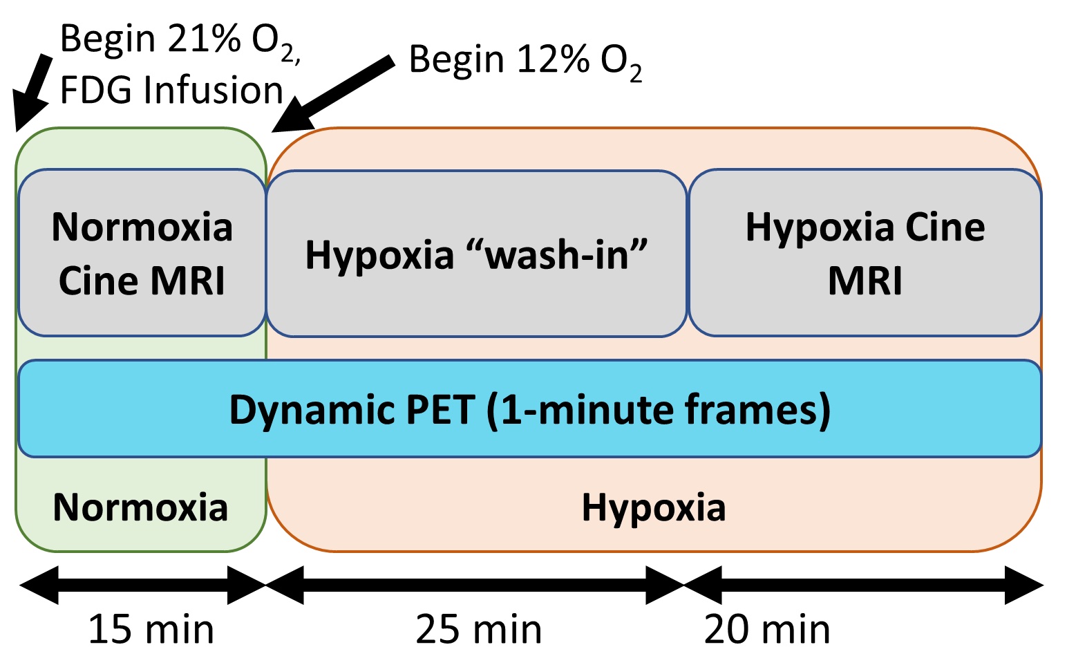
In clinical trials looking to study cardiac physiology in adults born extremely premature, my team is assessing pharmaceutical interventions (NCT03696758 and NCT04090866 on clinicaltrials.gov) and hypoxic gas as a stress test (NCT03245723). 
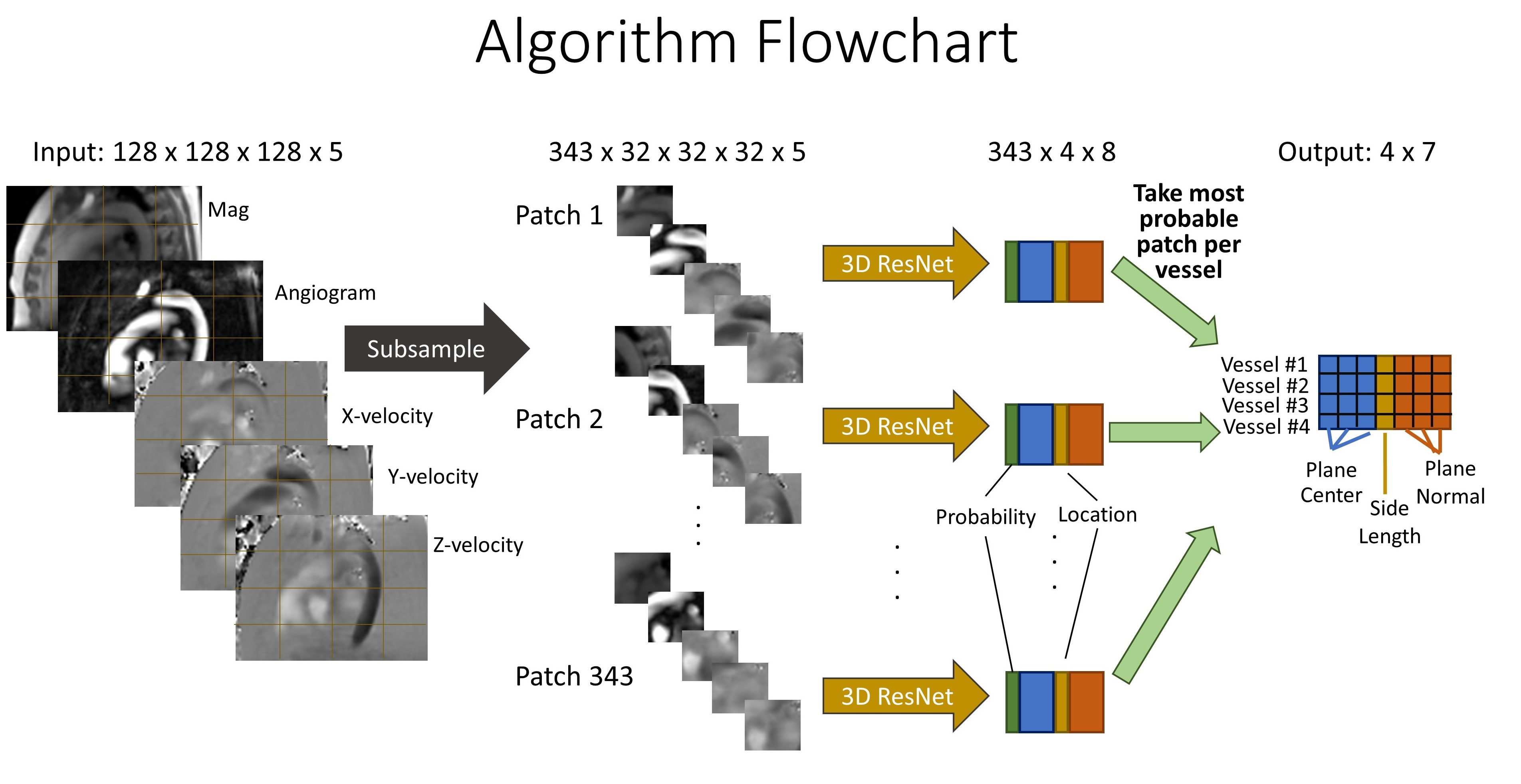
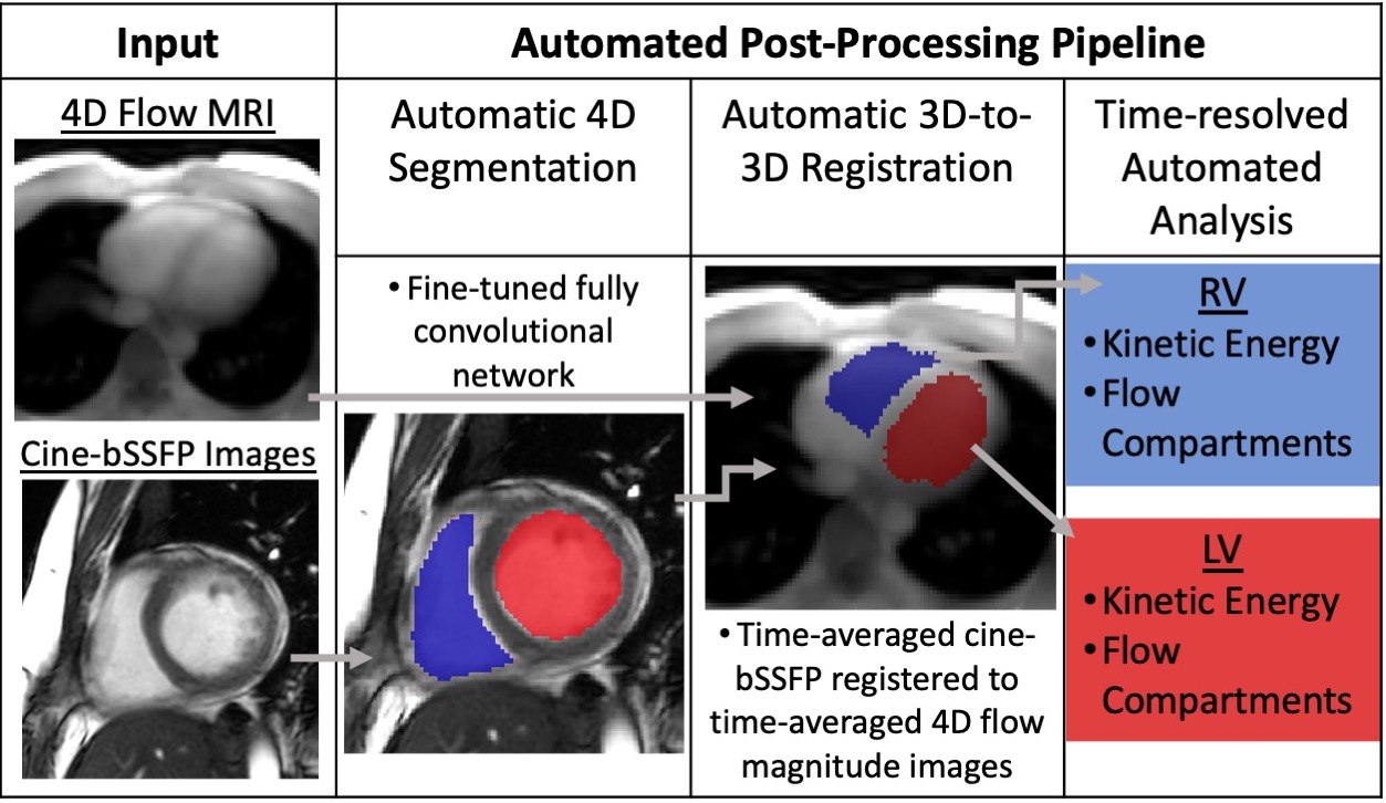
Throughout the projects I’ve taken the lead on image analysis methods for advanced cardiac MR images, writing custom code in Matlab and python to measure imaging biomarkers demonstrated in the literature. I’ve also led the statistical analysis and interpretation efforts pertaining to these and PET images, with help from clinicians and biostatisticians, resulting in 3 first-authored publications in the late stages of peer-review stemming from these trials. In the later 2 trials, I’ve also been involved in setting up the imaging protocols including MRI and PET acquisition parameters and data management.

Dual-Venc 4D Flow MRI Acquisition & Reconstruction

4D flow MRI uses the sensitivity of spins' phases to velocity and gradient first moments to map the 3-directional, time-resolved velocity field in a volumetric region in vivo. Traditional 4D flow MRI has only a narrow range of velocities it can be sensitive to, determined by the size of the gradient first moments. Dual-venc (DV) velocity encoding increases the sensitivity to slow flow while maintaining a high dynamic range of velocities that can be measured. The downside of this method is it requires longer scan times. To overcome this addition, I compared two undersampled radial trajectories in vitro for accelerated DV cardiac 4D flow imaging: 3D radial (PC-VIPR) versus stack of stars radial (PC-SOS), with benchtop particle imaging velocimetry serving as a reference standard. My paper on the comparison has been published in Magnetic Resonance in Medicine.

Real-time Cardiac bSSFP MRI Acquisition & Reconstruction

Cardiac MRI (CMR) is a powerful tool for assessing myocardial anatomy and function. Traditional breathheld 'cine' acquisitions take data from multiple heartbeats and combine them to reconstruct one composite heartbeat. ‘Real-time’ imaging instead of cine uses image acceleration to acquire data fast enough to reconstruct images with beat-to-beat cardiac dynamics, Thereby removing the need for breathholds and cardiac gating, enabling imaging of patients during an exercise stress test that cannot hold their breath. I employed aggressive undersampling with radial sampling, parallel imaging, and compressed sensing for real-time CMR during exercise and analyzed the performance of this approach in a numerical phantom and in a human volunteer.